

Journal of Creation 36(1):15–17, April 2022
Browse our latest digital issue Subscribe
Evolution of the neuron

The chasm that exists between nerve cells and their proposed evolutionary precursor cells has never been bridged by evidence, nor even by theoretical just-so stories. The problem of the evolutionary origin of the neuron is widely acknowledged by many evolutionists. The reasons why this gap exists, and will never be filled, are documented here. The first neuron must have had all of its basic parts assembled and integrated properly in order to function as a neuron.
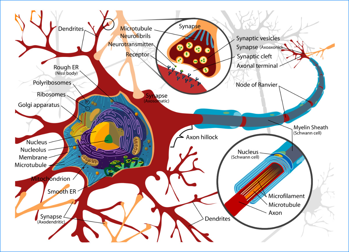
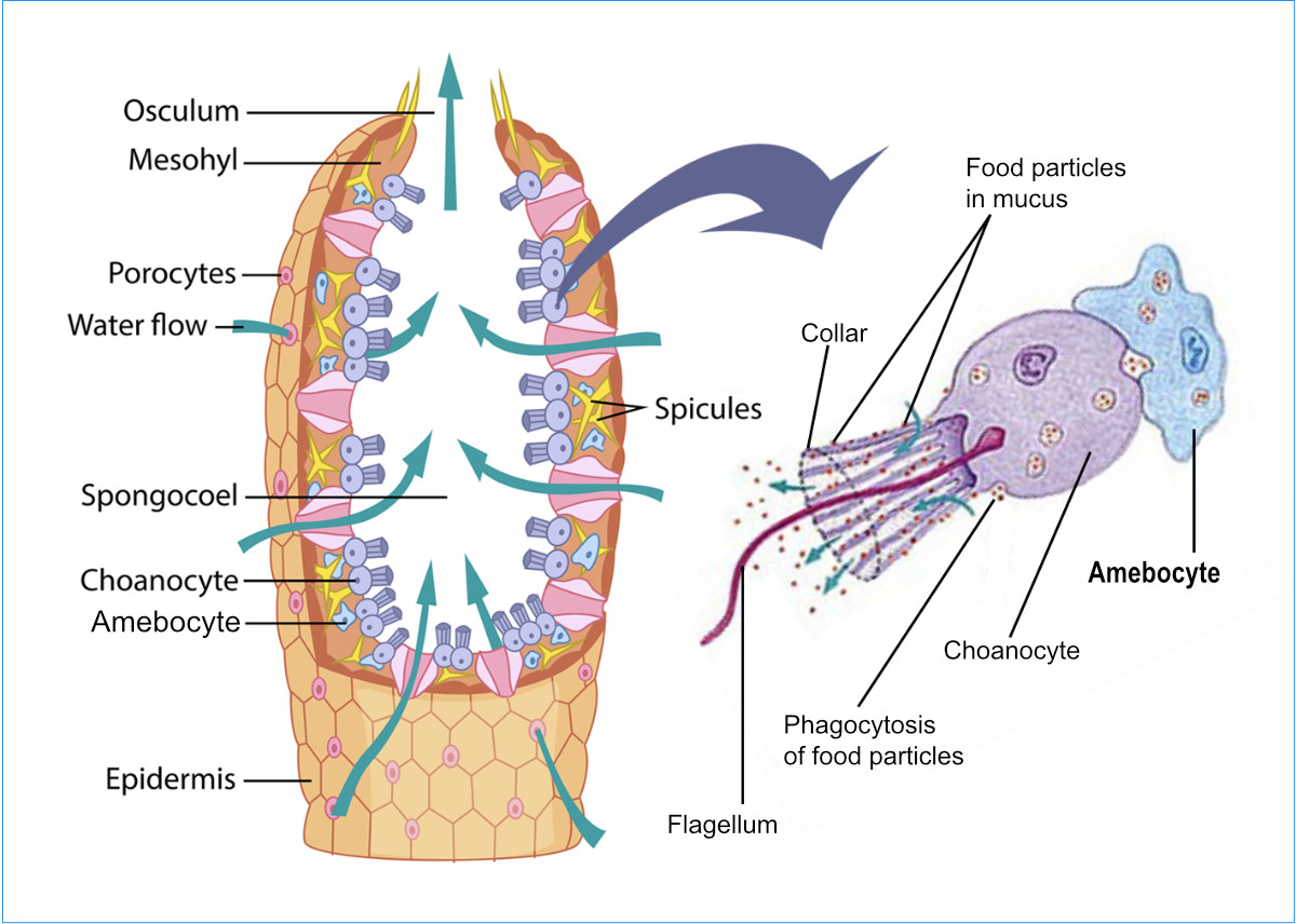
A neuron is another name for a nerve cell. It transmits information within an organism (for example, sensory information) to the brain. It communicates with other such cells by specialized connections, which are actually switches called ‘synapses’ (see figure 1). Nerve cells are employed in all animals except placozoa and sponges. Placozoa, the simplest existing non-parasitic metazoan, are small, marine, free-living multicellular organisms. Sponges (phylum Porifera) are multicellular metazoan filter feeders, the bodies of which are saturated with pores and channels, allowing water to circulate through them so they can absorb nutrients. As will be discussed, structures used by both of these animals have been proposed as supporting evolutionary ancestors of neurons.
In order to send messages, nerve cells employ a combination of electrical and chemical signals, which can be excitatory or inhibitory. The three neuron types are sensory neurons, motor neurons, and interneurons. Sensory neurons receive signals generated in the sensory organs (such as the eyes, ears, skin, olfactory organs, and taste buds) in response to light, sound, touch, pain, smell, and taste. They then transmit the information to the spinal cord or brain for processing and responding. Motor neurons receive signals from the brain and/or the spinal cord to regulate muscle contractions and glandular output. Interneurons connect neurons to other neurons.
Anatomy and physiology of neurons
Every neuron consists of a compact cell body called a soma, which, as is true of most cell types other than a few enucleated types (e.g. human red blood cells), has a nucleus. The nucleus contains DNA and part of the cellular machinery required to produce protein to repair and maintain the nerve cell. Most neurons have many dendrites, plus a long single axon (figure 1). Dendrites extend out a few hundred micrometres from the soma or cell body. The axon is the thin ‘cable’ along which the signal travels, and nerve fibres consist of bundles of axons. It leaves the soma at a swelling called the axon hillock. In humans, axons can be metres in length. At the farthest tip of the axons are terminals called synapses, where the neuron can transmit the signal across the synapse gap to another cell. The synapse regulates, by a system of chemical neurotransmitters, if the signal should cross to the next step in its journey (figure 1).
Evolution of the neuron
The first proposed evolutionary precursors to neurons are the cell types known as choanocytes or ‘collar cells’. Choanocytes are flagellated cells connected to a protoplasm collar located at the base of a flagellum (figure 2). They line the internal chambers of sponges. Their function is to move water into the sponge, where nutrients are then absorbed from the water. Choanocytes have a very different design to nerve cells but are proposed as the precursors of neurons because they have a flagellum, and because no better proposal exists. Nerve cells do not have a flagellum, but their axons look superficially like flagella. Note the contrast between the neuron in figure 1 and the choanocyte in figure 2.
One reason given for difficulty in documenting the evolution of neurons is the belief that soft tissue is not preserved in the fossil record. On the other hand, there is no shortage of living fossils, such as Cnidaria (including jellyfish), which, on the evolutionary timescale, “showed relatively little morphological change in the last 500 million years” since they first appeared in the fossil record.1 Darwinists argue without direct evidence that neurons must have evolved much farther back in time. In this case, time is used to explain away the lack of fossil evidence.
Another proposed precursor to neurons is the mesenchymal cell. These have cellular protrusions that resemble modern interneuron and motor neuron morphologies.2 Mesenchymal cells are undifferentiated stem cells that are able to develop into connective tissue, blood vessels, and lymphatic tissue cells, respectively. How they could have evolved into actual nerve cells is unknown. Stem cells are programmed to develop into certain cell types and will not develop into neurons unless the appropriate program exists and is triggered, and the environment is supportive of a neuron.
Neurons are irreducibly complex, so Stetka admits:
“We know neurons didn’t arrive in an instant. Instead, they evolved from relatively simple elaborations on earlier cell types and traits, maybe from epithelial cells, the cells that make up our skin; or from choanocytes, the early assemblers of animal life [emphasis added].”3
Assuming that a functional nerve cell could have evolved, the next step is for these nerve cells to evolve into nerve nets composed of neurons with neurites (a collective name for both axons and dendrites) in a mesh-like arrangement covering large parts of the animal body. This design is employed in modern ctenophores and cnidarians.4 The nerve net requires directions to assemble the net to function, involving a sensory system to receive information, and a muscle or other system to respond to the information. Until these systems are in place, the nerve net will be worse than useless and, at best, will just sit there and waste space and nutrients.
Nonetheless, the nerve net is considered one ‘hypothesis’ (among many) for the evolution of the central nervous system.4 The problem is that at this level evolutionists assume nerves have already evolved. Consequently, the focus of this review is the supposed evolution of the neurons themselves from some non-nerve cell. As Arendt writes: “Major questions in the evolution of neurons and nervous systems remain unsolved, such as the origin of the first neuron … .”4
Major problems for evolution
The major problem for evolution is that the neuron is one of the most complex cells in the body, and the gap between a neuron and all other cells is enormous (see figures 1 and 2).5 The human brain, containing some 100 billion neurons, which together form a complex network, has been called “the most complex object in the known universe”.6
Another problem for evolution is that, although all neurons have the same basic parts, many differences exist. Some axons are covered by a myelin sheath (see figure 1), composed of protein and lipids, in a manner similar to the insulation that surrounds electrical wires. The myelin sheath’s function is to speed up nerve transmission. Its deterioration, such as by an autoimmune disorder, causes debilitating diseases such as multiple sclerosis. These and other differences, such as the tissue types that develop into neurons in various animals, have forced some researchers to conclude: “Did neurons evolve more than once? Almost certainly.”1
Consider the following:
“Even if ctenophores and cnidarians are sister groups, with a neuron-carrying ancestor, some cnidarian neurons derive from endodermal cells rather than from epidermal cells, as is the norm, and the same epitheliomuscular cells in Hydra, a cnidarian, can be transformed into neurons by perturbing neurogenesis … . The multiple origins of neurons may, if fact, be why defining ‘neuron’ is so difficult, and why defining the origin of neurons is so complex.”1
Genetic comparisons, which were expected to solve the phylogeny of neuron evolution, have only made the problem worse:
“Does the feature’s absence in clade 2 mean that the feature was never present in the ancestors of clade 2, or was it present in clade 2’s ancestors but subsequently lost? A second phylogenomic problem is posed by convergent evolution (or ‘homoplasy’ in genetic terminology): a feature or a molecule that is present in two clades might have evolved independently in each clade. Both of these problems, secondary loss and homoplasy, confound the interpretation of evolutionary relationships.”7
Further complicating the evolutionary story is the conjecture that the “Three kinds of gated channels probably evolved independently.”7 For a nerve impulse to be carried forward, it must cross a gated channel between the axon and the next cell structure. Gated channels are molecules that form synaptic structures which control the messages’ travel from the sensory receptor across the neuron to the message receptor (the brain, for example).
The three kinds of gated channels are 1) the voltage-gated channel, 2) the stretch-gated channel, and 3) the ligand-gated channel.
The voltage-gated channel is a membrane that opens and closes in response to changes in membrane potential (voltage). In neurons, the sodium and potassium channels are examples of this type. The stretch-gated channels respond to membrane stress and are common in sensory cells. Lastly, ligand-gated channels are a group of trans-membrane ion-channel proteins which open to allow ions (including Na+, K+, Ca++, and/or Cl−) to pass through the membrane in response to a chemical messenger (i.e. a ligand), such as a neurotransmitter (figure 1).
This is another example of the issue:
“… whether neurons evolved only once or have different evolutionary origins—as suggested for example by the major differences in transmitter usage, synapse architecture and neuronal morphology observed in ctenophore, cnidarian and bilaterian nervous systems.”4
A non-evolutionary explanation accounts for this situation quite well. Each type and its claimed ancestors were separately designed; thus, the very implausible multiple evolution origin of the neuron and evolutionary-loss hypothesis are unnecessary.
Conclusion
“How did a structure as complex as our own brain ever evolve? … biologists have pondered this question since Charles Darwin.”7 And the fact is that, more than 150 years later, “The evolution of neurons and the nervous system is one of the remaining great mysteries of animal evolution.”4 A great chasm exists between the postulated precursor cells, the choanocytes, and the neurons that exist in every animal except placozoa and sponges. Nevertheless, much research has been expended to answer this question because in the study of the nervous system, “One of the most exciting questions [in evolution] is when and in what form the first neuron emerged … [emphasis in original].”4
References
References and notes
- Kristan, W.B. Jr, Early evolution of neurons, Current Biology 26:R937–R980, 2016; R954. Return to text.
- Arendt, D. et al., Evolution of neuronal types and families, Current Opinion in Neurobiology 56:144–152, 2019. Return to text.
- Stetka, B., A History of the Human Brain from the Sea Sponge to CRISPER: How our brain evolved, Timber Press, Portland, OR, p. 36, 2021. Return to text.
- Arendt, D. et al., ref. 2, p. 144. Return to text.
- Flatow, I., Decoding ‘the Most Complex Object in the Universe’, NPR, 14 June 2013, npr.org/2013/06/14/191614360/decoding-the-most-complex-object-in-the-universe. Return to text.
- New Scientist, How Your Brain Works: Inside the most complicated object in the known universe, London, UK, 2017. Return to text.
- Kristan, ref. 1, R949. Return to text.

Readers’ comments
Comments are automatically closed 14 days after publication.