

Journal of Creation 25(3):89–95, December 2011
Browse our latest digital issue Subscribe
Countering revisionism—part 1: Ernst Haeckel, fraud is proven
For more than a century, one of the foremost bastions of Darwinian evolution has been that embryos of different animals pass through a similar stage in which they resemble one another very closely. Although embryologists had long known this to be false, a bomb exploded in 1997 when an embryologist actually published real photos of embryos, showing many more differences than previously thought. The embarrassment to the evolutionary community was severe. But now a historian has made a serious attempt to rehabilitate Haeckel by revising both the history and the science around his claims.

Ernst Haeckel (1834–1919) was a professor of zoology and marine biologist, as well as a qualified medical doctor who was involved at the University of Jena during most of his academic lifetime. Besides his interests in biology, he was also a passionate artist who paid attention to many fine details in his artworks. His artwork was mainly about living creatures. But Haeckel is perhaps best known for his deception, using his wonderful talent as an artist combined with his authority as a scientist to convince people that Darwinian evolution is a fact. This specifically applies to sets of embryos which Haeckel drew and published in his very popular works Natürliche Schöpfungsgeschichte1 and Anthropogenie2. Ever since the publication of these sets, it has been controversial, and fellow scientists felt it was at best a misrepresentation of reality, at worst deceptive and fraudulent. (The latter was ultimately shown to be the case.)
Despite the controversy, textbook authors and teachers of evolutionary theory keep on using these diagrams, or versions of them,3 in order to convince students of evolutionary truth, even in the 21st century!4 In 1997, a ‘bomb’ exploded in the face of all those evolutionists who so fondly kept on using this evolutionary ‘icon’, when embryologist (and evolutionist) Dr Michael K. Richardson and his colleagues published a variety of real photographs of the relevant embryos.5 These drawings of Haeckel were later compared directly to the actual photos, and they were found to be far more different than everybody even thought. Richardson also published photographs of species additional to those which appeared in Haeckel’s popular embryo plates. This showed that Haeckel conveniently used those which tended to look more similar, while ignoring those which were different.
Although a minority of honest evolutionists have appreciated Richardson’s work, such as Stephen Jay Gould, Scott F. Gilbert (author of developmental biological books) and Paul Dombrowsky (a specialist in rhetoric), the embarrassment was just too severe and the iconic embryos too beloved among textbook authors to let things stay as they were. Robert John Richards, a professor of history at the University of Chicago, made a concerted attempt to rehabilitate not only the history around Haeckel, but also the very embryo sketches themselves. In 2008/9 Richards published a book and a paper in which he made some serious attempts to clear and clean up the name of his hero, Ernst Haeckel. My paper will look mainly at the works of Haeckel and the scientific issues around them, specifically set out in Richards’ paper named Haeckel’s Embryos: Fraud not proven.6 Where necessary, related issues will be discussed.
Michael Richardson and his co-workers’ photos of actual embryos had shown just how far Haeckel’s illustrations were from reality. It is thus no surprise that Robert Richards tries every possible thing to disprove Richardson and others’ work and critiques it as “logically mischievous, historically naive, and founded on highly misleading photography” (p. 148). His target is fully set on the photos of Richardson et al.
So just what exactly is technically wrong with Haeckel’s illustrations? What did Haeckel do in order to make his embryos look much more similar than they really are in general (and perhaps fish-like in particular)? From a fresh point of view, we can also find additional errors that have not previously been pointed out.
Technical errors with Haeckel’s illustrations
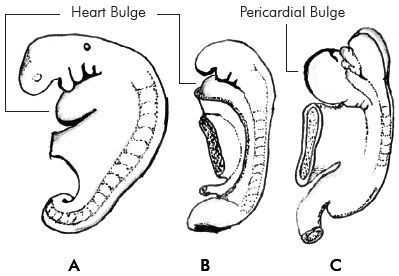
Heart Bulges
One of the first problems with the illustrations in the first row of Haeckel’s comparative embryo plates in his work Anthropogenie is that he drew many embryos, including the human and chick embryos, without either pericardial or heart bulges, where they possess these in reality. In humans, the cardiovascular system is one of the first entities to develop in the early embryo. This is so because the growing embryo needs a constant supply of oxygen, and nutrients. Very early in embryonic development, diffusion becomes insufficient for oxygen supply.7 So from even as early as 25 days old, the human embryo already displays a clear pericardial bulge, soon becoming a heart bulge (figure 1). In the earliest row of illustrations in Anthropogenie (figure 4 below, first row), Haeckel’s human sketches lack these heart bulges. This is the case not only of the above mentioned work, but also other works, including the late editions (for example the 12th edition) of Natürliche Schöpfungsgeschichte, and a book drawn up as a collection of popular lectures called Last Words on Evolution.8 In the 4th and 5th editions of Antropogenie, the error keeps on being repeated. We should at this stage make the important observation that Haeckel was a fully qualified medical doctor, and he was thus well acquainted with human biology. So he is without excuse for misrepresenting human physiology in this way.
The same principle applies for some other animal groups as well, specifically the chick embryo. In the chick, the blood starts circulating at the 16-somite9 stage (about 36–37 hours old) where the ventricle is already visible. A bulge (consisting of the ventricle and atrium) becomes clearly visible at the 19-somite stage (about 43 hours old) and is even more pronounced at the 26-somite stage (about 51–53 hours old).10 Haeckel’s chick embryo in the 1st to 3rd editions of Anthropogenie matches best the last-mentioned stage, except that no sign of the cardiovascular system is visible in these editions of the plates.
The problem is not just for the human and chick embryos. Some other classes of vertebrates have the same problem, whilst other classes of vertebrates, like certain species of fishes and amphibians, may not display heart bulges at all (at least visibly). This is the first clear distortion by Haeckel in order to make these embryos look more similar.
Limb buds in embryos
Another thing which seemed to have surfaced as erroneous with Haeckel’s illustrations is the fact that embryos lack limb buds at certain places where they should show them. But first, biologist Scott Gilbert draws our attention to something important:
“Interestingly, there was some discussion as to what exactly this stage was (Richardson 1995). This conserved stage was sometimes considered the neurula stage (Wolpert 1991), the ‘pharyngula’ stage (characterized by the branchial arches; Ballard 1981), the tailbud stage (Slack et al. 1993), or the stages between those of headfold and tailbud (Duboule 1994).”11
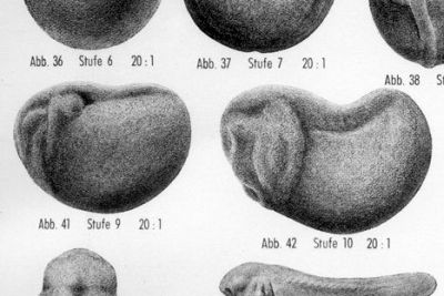
Gilbert goes on to explain that heterochrony (the phenomenon of different timing in the appearance of structures) is another problem in general. Specifically, at whatever stage is selected for comparison, some species’ embryos will display limb buds, whilst others may not at all. This is actually seen in more of the photos which Richardson et al. have published.12 Naturally, the question arises whether Haeckel himself was aware of this. The answer is a remarkable ‘yes’, at least for two reasons. First, Gilbert further points out: “Interestingly, this knowledge [of heterochrony] appears to be ‘old hat’ among German biologists.”11 Second and more importantly, though, there seems to be clear evidence that Haeckel purposely removed limb buds from embryo drawings of his sources, in order to make them look more similar. In a correspondence to the editor of Nature,13 Richardson and Keuck explain, and show pictures of, how Haeckel purposefully removed the limb buds from an echidna-embryo drawing. His source was a work14 of Richard Semon, who used the original drawing in at least two works. Haeckel himself used this limbless drawing in at least two places as well, the 5th edition of his Anthropogenie and the late editions (such as the 12th) of Natürliche Schöpfungsgeschichte. We can thus clearly see how Haeckel intentionally distorted embryo drawings in order to make them look more similar (figure 2).

Richards is aware of this paper, and tries to make yet another excuse for Haeckel’s deliberate deception.15 He tries to argue that Haeckel adapted the embryo drawing for an earlier stage of development than the one in which Semon’s illustration was. But if that were the case, then Haeckel should have also adapted other morphological features, which he did not (as Richards admits in his book, thinking he is doing Haeckel a favour). The more we go back in the early embryonic stages from the relevant point, the lower the somite number would have needed to be (in fact, another way of measuring the stage in which an early embryo is, is referring to the amount of somites present in the embryo). Also, the pharyngeal arches would be less pronounced, and other features more generalized (as per the embryonic principles of Karl Ernst von Baer (1792–1876), with whom Haeckel was also familiar). Haeckel must have known all the above. He was very familiar with early embryonic development, and even coined what we know as ‘Gastraea Theory’ (describing even earlier embryonic development). Haeckel adapted none of these features, especially not the pharyngeal arches, which he so fondly referred to as ‘gills/ gill slits’. Richards’ excuse simply falls flat, and turns out to be a convenient ad hoc theory.
If the above is not enough, Richardson and Keuck found yet another example of Haeckel removing limb buds.16 This time it was from a tuatara (Sphenodon punctatus from New Zealand) embryo which he seemed to have copied from Arthur Dendy17 (1865–1925). Again, there can be no excuse for this deliberate misrepresentation.
To come back to the original point, we know from history, as well as photography and theoretical knowledge, that the problem of limb buds is a general one, and not just limited to the echidna and tuatara. And second, that Haeckel seemed to deliberately have removed these limb buds. Richardson and Keuck found several other examples of Haeckel’s removing limb buds from original sources as well.18 The error is also often repeated in modern textbook drawings of the iconic embryos.
Yolk and photography
In order to save Haeckel from the obviously clear falsehood(s) demonstrated by Richardson et al.’s photos, Richards makes a full-fledged attack on the photography by desperately trying to make an argument regarding the yolk. But first it should be firmly noted just how differently yolk is incorporated in the development of embryos of different types of animals. One insightful source mentions, for instance, the following differences:
“Many animals (e.g. many insects, octopuses, fish, reptiles, marsupial mammals) use yolk sacs to feed the embryo … But there are also a number of animal groups (e.g. nematodes, sea urchins and almost all amphibians) that do not develop a yolk sac. In such organisms, the yolk is less conspicuous and is perhaps best defined as the nutritional reserves provided by the mother, including yolk platelets, fat droplets and glycogen [emphasis added].”19
Even among mammals, there exists noticeable variety:
“Monotremes, such as the platypus, and marsupials, such as kangaroos, have large, yolky eggs [since they actually lay eggs]. Placental mammals, by contrast, have small eggs without yolk platelets. … Even placental mammals still form extraembryonic yolk sacs and these are by no means useless vestiges [emphasis added].”20
So we can safely conclude that the development of yolk is yet another important difference in embryonic development. But Robert Richards makes specific claims about the yolk (which he does not really seem to understand anyway):
“… several (but not all) of the photographed embryos retain the attached yolk sack and other maternal material; this exaggerates their differences from Haeckel’s images … . The bulge of the salamander is not part of the embryo; rather, it is the yolk sack, as is the case for the fish and the human embryos (though not for the chick and the rabbit, from which the yolk sacks [sic] have been removed) … .”21
First of all, in the original Richardson paper we are told that:
“The extra-embryonic membranes were either missing or were removed by us. However the allantois was preserved where present.”22
So this team of biologists actually were careful with extra-embryonic materials. And contrary to what this historian (Richards) says, the bulge (not a typical yolk sac, in the biological sense of the word) of the salamander is part of, and attached to, the body of the embryo, unlike human embryos where a yolk sac is outside of the embryo itself. In many other species as well, it would be impossible to separate the yolk from the body of the embryos without doing violence to the structure of the embryo, and misrepresenting it. So, exactly contrary to what Richards says, by removing these properties, the similarities are exaggerated between these embryos. This is thus yet another embryonic feature which Haeckel distorted, as we have seen. Moreover; the very usage of a salamander embryo as representative of the class of amphibians speaks of convenient selective reporting by Haeckel. Frogs and toads represent the overwhelming majority of the class of amphibians, and their embryos completely break the common visual similarity pattern at the tailbud stage of embryogenesis—refer to figure 3 to see just how pattern-breaking frogs in general are.
Returning to the yolk issue, the development of yolk adds greatly to the variety in the development and appearance of different embryos of species. Richards’s arguments to discredit the photography of Richardson et al., and to salvage Haeckel, thus fail. We must also point out that yolk cannot be written off as irrelevant to embryonic development. The way embryos of different species undergo cleavage is much determined by the yolk. It also determines how later stages follow. A lot of yolk means the embryo goes directly to a little adult, while little yolk means that it develops into various larval stages.23

Richards then took some of Dr Richardson et al.’s photographs which appeared in Elizabeth Pennisi’s article24 in Science (also widely used by creationists to expose the myth25,26) and re-engineers them according to what he thinks they should have looked like (figure 4). Yet in doing so, he too, astoundingly enough, produces exactly the same errors as did Haeckel himself! The most important of these is the heart bulge of the human embryo, which is completely removed in Richards’ re-engineering. Such a removal cannot by any means be justified. Other tactics include straightening out the chick embryo’s torsion and flexion (literally ‘twisting’ the body), which is significant in the development, as well as the reengineering of the salamander body, in order to get rid of the bulge. As for Haeckel’s own distortion of the torsion and flexion, the excuse is dished up that those processes occur at a somewhat later stage of development. But comparing other features of Haeckel’s chick embryo to the relevant literature reveals this as yet another fact-free excuse.27 Since Haeckel’s chick embryo had both visible developing eyes and pharyngeal arches, this cannot be true, and embryos with no flexion and torsion are clearly in a too-early stage to match that of Haeckel. Richardson et al. also carefully picked their chick embryo to be at the correct developmental stage, so Richards’ doctored chick embryo does not exist in nature.
Issues with editions of Anthropogenie
Richards whines about the set of embryos that was used for comparison to actual photos by Michael Richardson et al. Richardson had used the illustrations from the 1874 (first) edition of Anthropogenie. Richards implies that it was unfair of Richardson to use these, and tells us:
“In the subsequent editions, the images grew ever more refined, so that even by the 4th edition (1891), the differences among them became more pronounced … .”28
But this gives an entirely wrong impression of gradual improvement. Richards fails to mention that the original sketches are found even in the 3rd edition! Seeing the fact that the book only went through five editions, it represents the majority of them. Using the 1st to 3rd editions’ drawings is further justified, because those sketches were used in countless textbooks ever since. A major and important example is George Romanes’ book Darwin and After Darwin.29 This book gave authors30 the option of citing these embryo drawings from Romanes, thus ‘sanitizing’ them from Haeckel’s name.31 The only differences between Romanes’ version of them and Haeckel’s are that Romanes removed annotations and used a white background, where Haeckel used a black or dark background, but they are structurally identical. Many evolutionists argue that we should make a distinction between Haeckel and Romanes, but there is no valid reason for doing so. Some authors32 shamelessly use Haeckel’s 1874 sketches directly! Recently (2010) these dubious sketches were even on the front page of the prestigious journal Nature.33 Furthermore, the tradition of using the same animal sequence (we cannot even say species in this regard because of over-generalization) for comparison in textbooks, as did Haeckel in his original Anthropogenie sketches, has carried on, even in the 21st century. The sequence is: fish, salamander, tortoise, chick, hog, rabbit, calf and human. Sometimes authors conveniently leave out those which they know don’t fit. Hickman et al., in their Integrated Principles of Zoology (2008), and Sylvia S. Mader’s Biology (10th edition), are good cases in point of authors still using the 1874 Haeckelian sequence and selection of animals, which is conveniently selective. Both of these above editions were recent when this paper was written.
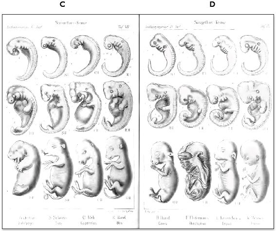
After the above quote, Richards refers us to embryo drawings from the 4th edition of Anthropogenie, which is supposed to show us that Haeckel pronounced the differences much more, thus improving them. But it is precisely here that we find probably the most dubious aspect of Richards’ paper. To explain, in the 4th edition Haeckel presented not embryos of 8 species of animals (like in the 1st to 3rd editions), but 14 species of embryos in four plates reaching over four pages (or two double pages). To illustrate his point, Richards gave his readers what seems to be one of the double-page illustrations, but Richards has put the wrong plates together, giving us the two right-hand ones (‘B’ and ‘D’ in figures 5 and 6, not belonging together on the same page) without giving the slightest hint about it to his readers! Of course the differences would look more pronounced! See figures 5 and 6 to understand why. Such disingenuous selective reporting hardly shows either Haeckel’s or Richards’ intentions in a good light.
As one can expect, the more Richards’ work is becoming known among members in the evolutionary community, the more anti-creationists are eager to use this as an antithesis for creationist books and work. Shockingly, but not surprisingly, the pretentiously named National Center for Science Education has already picked this paper up,34 quoting Richards on several places and using Richards’ wrong illustrations just as they are.35 Creationists should be ready to point out this extremely sloppy, not to say dishonest, scholarship.
Richards does not show us the additionally added embryo drawings of the 5th (and latest) edition of Anthropogenie in his paper, only in his book. But might we point out that amongst these additional embryo sketches in the 5th edition, the limbless echidna as well as the tuatara are to be found, once again suggesting deliberate inaccuracies. The human embryo is also still without its heart bulge in the 5th edition.
 |
 |
Issue with sizes of embryos
Another complaint being made against Richardson et al. is one about size. Richards tells us:
“… Richardson suggested that Haeckel ‘fudged the scale’ of the embryos, even though there was a tenfold difference in magnitude among them. Haeckel, however, quite explicitly stated in the caption to his illustration that he reduced all of the images to the same size to facilitate structural comparisons … .”36
Haeckel did indeed mention, in brackets in the explanatory descriptions of the plates of Anthropogenie, that he reduced all the embryo illustrations to the same size for comparison. So, this may be one of the only valid objections by Richards. However, complaints about sizes of some embryo comparisons in earlier editions of Natürliche Schöpfungsgeschichte may still not have been too unreasonable, since some of the information about sizes was not quite correct. With the Anthropogenie sketches, however, we must let Haeckel off the hook with this one, which was never one of the most serious complaints to begin with.
Other issues
Richards writes as if everyone in past and present is so extremely unfair to ‘poor old’ Haeckel. He makes points on which we must both agree and disagree. We are told:
“Parity of reasoning should logically have required another conclusion: if the indictment of fraud should be made against Haeckel because of too-similar images, then it ought to be brought also against His and the many modern embryologists whom Richardson and his colleagues cited, since they, too, supposed a phylotypic stage in embryogenesis [then citing the embryologists also cited by Gilbert, ref. 7].
“They maintained that not only did Haeckel’s images misrepresent the actual state of embryos but so did those of Wilhelm His, perhaps the most famous embryologist of his day and Haeckel’s bitter enemy. His, they contended, also exaggerated the similarities of embryos and ignored their differences.”37
We can agree that it is not only Haeckel that should be held responsible. Many textbook authors who so shamelessly use too similar drawings and repeat errors are also guilty, although in some cases only of trusting previous works, not intentional deception. But the problem with Richards’ reproach to Richardson and other authors who then wrote articles about it in other journals (following Richardson’s original paper) is that they do blame other people besides Haeckel! Paul Dombrowsy38 (an expert in rhetoric), Stephen Jay Gould,39 and even Richardson, in a letter to Gould40 (which got published), for instance, do hold many others responsible.
Furthermore, the charge against Wilhelm His is most odd, because Richardson and Keuck tell us exactly the opposite about Wilhelm His’s work:
“Haeckel’s young embryos look similar, whereas His’s look different.”13
They explain that whereas Haeckel’s embryo drawings stressed the similarities, His’s embryos tended to stress the differences. Although Richardson and Keuck feel that His’s embryos were not always accurate either, they point out that the issue becomes a question of intent. They haven’t found evidence that His deliberately distorted his embryos, whereas with Haeckel, they clearly have (by tracing Haeckel’s sources in this instance. See the removed limbs of the echidna, earlier). Yet this very paper (of Richardson and Keuck) is referred to and discussed in Richards’ book (in regards to the limb buds)! Why does he not inform his readers about this aspect in his paper?
Conclusion
The photographs of Dr Michael Richardson et al. are as valid as when they were first published. But creationists will now have to rely on more than just the visual illustration of these powerful photographs and also be capable of explaining why Haeckel’s original work is based on ideology and dishonesty. R.J. Richards’ attempts fall flat under closer investigation, and his scholarship is often extremely sloppy, and does not represent the facts accurately. The impression is frequently given that everybody (creationists and evolutionists) were just so unfair to ‘poor old Haeckel’, but this is not the case. On investigating Haeckel’s illustrations technically, it becomes clear just how many things Haeckel distorted in the embryo illustrations. His dishonesty can thus not be denied.
Finally, after having looked closer at some of the issues at hand, it is reasonable to maintain the position that Haeckel’s fraud is proven.
Acknowledgements
The author would like to thank Jonathan Sarfati and Daniel Davidson for their handy discussions and suggestions on this issue.
References
- Haeckel, H., Natürliche Schöpfungsgeschichte, G. Reimer, Berlin, 1868. Literally ‘Natural History of Creation’; English title: The History of Creation, or the Development of the Earth and its Inhabitants by the Action of Natural Causes: A popular exposition of the doctrine of evolution in general, and that of Darwin, Goethe, and Lamarck in particular, First English edition translated by Prof. Ray Lankester, Fellow of Exeter College, Oxford, 1876. Return to text.
- Haeckel, E., Anthropogenie oder Entwickelungsgeschichte des Menschen, Engelmann, Leipzig, 1874. Literally ‘Anthropogeny or History of Evolution of Mankind’, English title: The Evolution of Man—A popular scientific study, Translated from the 5th (enlarged) edition by Joseph McCabe (1867–1955), a vocal apostate and leading atheopathic campaigner, 1905. Return to text.
- See, for example, some documentation in: Luskin, C., What do modern textbooks really say about Haeckel’s embryos? Discovery Institute, March 2007, www.discovery.org/a/3935#text1, accessed 22 November 2010. Return to text.
- See, for example, this study of Texan textbooks in Luskin, C., Haeckel’s Embryo Drawings Make Cameos in Proposed Texas Instructional Materials, Evolution News and Views, 17 June 2011, www.evolutionnews.org/2011/06/haeckels_embryos_make_multiple047321.html, accessed 18 July 2011. Return to text.
- Richardson, M.K., Hanken, J., Gooneratne, M.L., Pieau, C., Raynaud, A., Selwood, L. and Wright, G.M., There is no highly conserved embryonic stage in the vertebrates: implications for current theories of evolution and development, Anatomy and Embryology 196(2):91–106, 1997. Return to text.
- Richards, R.J., Haeckel’s embryos: fraud not proven, Biology and Philosophy 24:147–154, 2009 | DOI 10.1007/s10539-008-9140-z; Published online: November 2008. Return to text.
- Mitchel, B. and Sharma, R., Embryology: An illustrated Color Text, Elsevier, Churchill Livingstone, p. 26, 2005. Return to text.
- Haeckel, E., Last Words on Evolution: A popular retrospect and summary, Translated from the 2nd ed. by Joseph McCabe; Peter Eckler, New York, 1905. Return to text.
- A somite (from Greek σῶμα soma = body) is a division of an animal body, formerly called a ‘primitive segment’. Return to text.
- This is neatly illustrated and annotated by fig. A-24 of Carlson, B.M., Patten’s Foundations of Embryology, 4th ed., McGraw-Hill, Columbus, OH, p. 566, 1981. Return to text.
- Gilbert, S.F., Haeckel and the vertebrate archetype; in: DevBio—A Companion to Developmental Biology, 8th ed., Sinauer Associates, Sunderland, MA; http://8e.devbio.com/article.php?id=242, accessed 18 January 2011. Return to text.
- For instance, figure 3 in DevBio, ref. 11.; Visible online at: http://8e.devbio.com/image.php?id=490, accessed 18 January 2011. Return to text.
- Richardson, M.K. and Keuck, G., A question of intent: when is a ‘schematic’ illustration a fraud? Correspondence to Nature 410:144, 2001. Return to text.
- Semon, R., Zoologische Forschungsreisen in Australien und dem malayischen Archipel, Fischer, Jena, Germany, 1894, as pointed out by Richardson and Keuck, ref. 13. Return to text.
- Richards, R.J., The Tragic Sense of Life: Ernst Haeckel and the Struggle Over Evolutionary Thought, University of Chicago Press, p. 336, 2008. Return to text.
- Richardson, M.K. and Keuck, G., Haeckel’s ABC of evolution and development, Biol. Rev. 77:509, 513, 2002, DOI: 10.1017} S1464793102005948. Return to text.
- Dendy, A., Outlines of the development of the tuatara, Sphenodon (Hatteria punctatus), Quarterly Journal of Microscopical Science 42, 1899, as cited by Richardson and Keuck, ref. 16. Return to text.
- Richardson and Keuck, ref. 16, pp. 512–514. Return to text.
- Jorgensen, P., Quick guide: yolk, Current Biology 18(3):R103, 2008. Return to text.
- Jorgensen, ref. 19, p. R104. Return to text.
- Richards, ref. 6, pp. 150–151. Return to text.
- Richardson et al., ref. 5, p. 95. Return to text.
- Hickman, C.P., Roberts, L.S., Keen, S.L., Larson, A., I’Anson, H. and Eisenhour, D.J., Integrated Principles of Zoology, 14th ed., McGraw-Hill, Columbus, OH, p. 163, 2008. Return to text.
- Pennisi, E., Haeckel’s embryos: fraud rediscovered, Science 277(5331):1435, 1997. Return to text.
- Grigg, R., Fraud rediscovered, Creation 20(2):49–51, 1998; creation.com/fraud. Return to text.
- Sarfati, J., Refuting Evolution, ch. 6, Creation Book Publishers, 2008. Return to text.
- Carlson, B.M., Patten’s Foundations of Embryology, 4th ed., McGraw-Hill, Columbus, OH, 1981; p. 566 figure A-24, as well as more explanation on page p. 569. Return to text.
- Richards, ref. 6, p. 150. Return to text.
- Romanes, G.J., Darwin, and After Darwin—An Exposition of the Darwinian Theory and a Discussion of Post-Darwinian Questions, Longman’s Green and Co., London, 1892. Return to text.
- For example, like Futyuma, D., Evolutionary Biology, 3rd ed., Sinauer Associates, Sutherland, MA, 1998. Return to text.
- As also pointed out by Gilbert, ref. 11. Return to text.
- For example, Gerhart, J. and Kirschner, M., Cell, Embryos and Evolution. Toward a Cellular and Developmental Understanding of Phenotypic Variation and Evolutionary Adaptability, Blackwell Science, Malden, MA. 1997. Return to text.
- Front page of Nature 468(7325), 2010. Return to text.
- Anon., Accuracy in embryo illustrations, 2008, on the NCSE’s website: http://ncse.com/creationism/analysis/exaggerations, accessed 20 January 2011. Return to text.
- Anon., Michael Richardson’s photographs, on the NCSE’s website: http://ncse.com/book/export/html/2320, accessed 1 July 2011. Return to text.
- Richards, ref. 6, pp. 149–150. Return to text.
- Richards, ref. 6, p. 149. Return to text.
- Dombrowsky, P., Ernst Haeckel’s controversial visual rhetoric, Technical Communication Quarterly 12(3), Summer 2003. Return to text.
- Gould, S.J., Abscheulich! (Atrocious!), Natural History 109(2), March 2000. Return to text.
- Published in Gould, ref. 39. Return to text.
- Langman, J., Medical Embryology, 3rd ed., The Williams and Wilkins Company, Baltimore, MD, 1975. Return to text.
- Illustration from Semon, R., In the Australian Bush and on the coast of the Coral Sea, Macmillan and Co., Limited London, p. 162, 1899. This echidna picture was also used in Semon, ref. 14, which Richardson and Keuck used for their exposé. Return to text.


Readers’ comments
Comments are automatically closed 14 days after publication.